Abdominal Anatomy - Abdominal anatomy, 1839 artwork - Stock Image - C022/3696 ...

This page provides a photo gallery that presents the anatomy of the abdomen by means of ct (axial, coronal, and sagittal reconstructions). • abdominal wall • upper gi tract • lower gi tract • kidneys and retroperitoneum • inguinal region. The linea semilunaris (open arrow); Anatomy of the abdominal wall, inguinal region & hernias, hernias, gut and peritoneal cavity. Identify some abdominal pathology on medical images. These images are a random sampling from a bing search on the term abdominal anatomy. click on the image (or right click) to open the source website in a new browser window. The above lines intersect and divide the abdomen into nine regions (clockwise from the top) Learn about abdominal organs anatomy with free interactive flashcards. 5 name the nine abdominal regions and their main contents. Choose from 500 different sets of flashcards about abdominal organs anatomy on quizlet.
Understanding abdominal anatomy and physiology is essential to understanding the human body as a whole. You will learn the anatomical basis of pain and how to apply this knowledge in the diagnostic process. Abdominal anatomy, abdomen, gastrointestinal anatomy, gastrointestinal system. Laterally by the midaxillary line; Become familiar with the anatomical divisions by exploring the world's most advanced 3d anatomy platform in complete anatomy. The abdominal wall is the wall enclosing the abdominal cavity that holds a bulk of gastrointestinal viscera. Learn about abdominal organs anatomy with free interactive flashcards. Who better to review abdominal anatomy with, than an experienced expert? Divided into 9 regions by two vertical and two horizontal imaginary planes. Identify abdominal anatomical structures in a variety of medical imaging platforms.
![]()
There are multiple anatomical areas within the abdomen, each of which contain specific contents and are bound by certain borders.
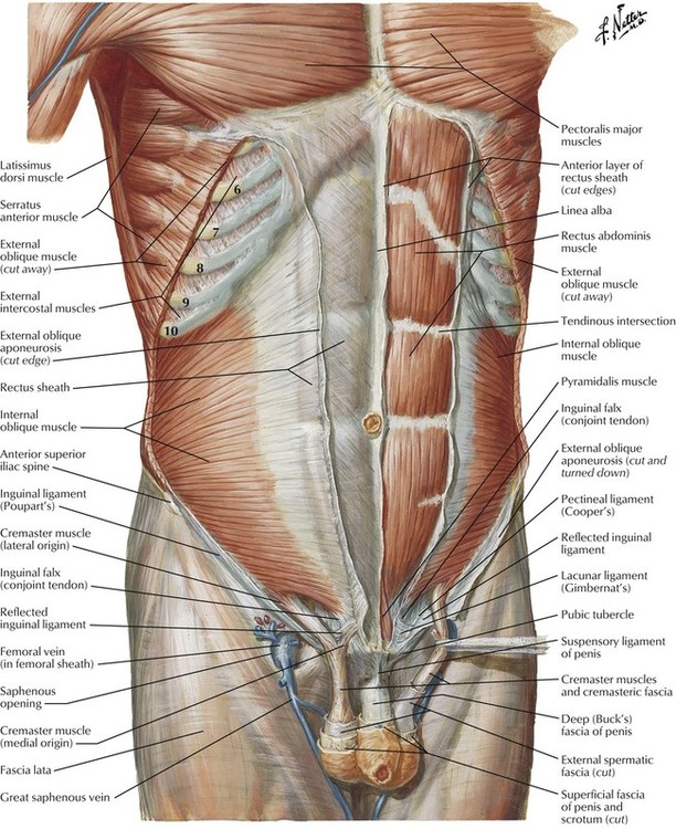
Abdominal cavity, largest hollow space of the body. Who better to review abdominal anatomy with, than an experienced expert? Mr anatomy of the abdominal wall demonstrating the three flat muscles (short arrow); Understanding abdominal anatomy and physiology is essential to understanding the human body as a whole. The abdominal divisions should be used in conjunction with other diagnostic approaches in order to accurately diagnose a patient's condition. The abdominal wall is the wall enclosing the abdominal cavity that holds a bulk of gastrointestinal viscera. The linea alba (open arrowhead); This muscle forms the anterior and lateral abdominal wall. 6 write the origin, insertion and nerve supply of muscles of anterior abdominal wall. The anterolateral abdominal wall formed of 4 layer skin, fascia, muscles, and peritoneum. The transversus abdominis muscle is the deepest of the abdominal muscles, lying internally to the internal abdominal obliques. Simple, easy notes for quick revision of important questions.
We created an anatomical atlas of abdominal and pelvic ct which is an interactive tool for studying the conventional anatomy of the normal structures based on a multidetector computed tomography. Identify abdominal anatomical structures in a variety of medical imaging platforms. Radiology basics of abdominal ct anatomy with annotated coronal images and scrollable axial images to help medical students and junior doctors learning anatomy. Anatomy of the abdominal wall, inguinal region & hernias, hernias, gut and peritoneal cavity. The abdominal region is supported by the anterior and posterior abdominal wall that supports the viscera and maintains the posture where there's no bony support. Demonstrate comprehension of core abdominal anatomy.

You will learn the anatomical basis of pain and how to apply this knowledge in the diagnostic process.
Simple, easy notes for quick revision of important questions. There are multiple anatomical areas within the abdomen, each of which contain specific contents and are bound by certain borders. Therefore, a firm grasp of abdominal anatomy is necessary to effectively diagnose and treat patients. In order to find the right training and to perform the exercises properly, it is important to know what are the abdominal muscles. We created an anatomical atlas of abdominal and pelvic ct which is an interactive tool for studying the conventional anatomy of the normal structures based on a multidetector computed tomography. Abdominal cavity, largest hollow space of the body. Identify some abdominal pathology on medical images. Become familiar with the anatomical divisions by exploring the world's most advanced 3d anatomy platform in complete anatomy. • in this module, we will explore basic abdominal anatomy identifiable with common imaging modalities. We'll identify as many organs as we can, see how they fit into. Two layers in abdomenfatty superficial layer (camper's fascia)deeper membranous layer (scarper's fascia). Windham was previously a surgical oncologist in the sarcoma program of the h. The epigastric vessels (long arrow); • the abdomen consists of:
There are multiple anatomical areas within the abdomen, each of which contain specific contents and are bound by certain borders. The epigastric vessels (long arrow); This page provides a photo gallery that presents the anatomy of the abdomen by means of ct (axial, coronal, and sagittal reconstructions). In order to find the right training and to perform the exercises properly, it is important to know what are the abdominal muscles. The transversus abdominis muscle is the deepest of the abdominal muscles, lying internally to the internal abdominal obliques. Compare and contrast the different medical imaging modalities presented in the tutorials. Anatomy of the abdominal wall, inguinal region & hernias, hernias, gut and peritoneal cavity. Divided into 9 regions by two vertical and two horizontal imaginary planes. Laterally by the midaxillary line;
These images are a random sampling from a bing search on the term abdominal anatomy. click on the image (or right click) to open the source website in a new browser window.
5 name the nine abdominal regions and their main contents. And inferiorly by the symphysis pubis, pubic tubercle, inguinal ligament, anterior superior iliac spine, and. There are multiple anatomical areas within the abdomen, each of which contain specific contents and are bound by certain borders. Sciency root words make anatomical parts harder to memorize. Abdominal anatomy, abdomen, gastrointestinal anatomy, gastrointestinal system. Learn about abdominal organs anatomy with free interactive flashcards. Abdominal cavity, largest hollow space of the body. The anterolateral abdominal wall formed of 4 layer skin, fascia, muscles, and peritoneum. Its upper boundary is the diaphragm, a sheet of muscle and connective tissue that separates it the abdominal organs are supported and protected by the bones of the pelvis and ribcage and are covered by the greater omentum, a fold of peritoneum. The quadratus lumborum muscle (black arrow). Divided into 9 regions by two vertical and two horizontal imaginary planes. Who better to review abdominal anatomy with, than an experienced expert? We'll identify as many organs as we can, see how they fit into. Abdominal surface anatomy can be described when viewed from in front of the abdomen in 2 ways:

Windham was previously a surgical oncologist in the sarcoma program of the h.

• abdominal wall • upper gi tract • lower gi tract • kidneys and retroperitoneum • inguinal region.

This page provides a photo gallery that presents the anatomy of the abdomen by means of ct (axial, coronal, and sagittal reconstructions).

The anterolateral abdominal wall formed of 4 layer skin, fascia, muscles, and peritoneum.

Abdominal cavity, largest hollow space of the body.

Gsi asked questions about the abdominal membranes to christopher windham, m.d.

You will learn the anatomical basis of pain and how to apply this knowledge in the diagnostic process.
 to open the source website in a new browser window.")
The abdominal divisions should be used in conjunction with other diagnostic approaches in order to accurately diagnose a patient's condition.
deeper membranous layer (scarper's fascia).")
6 write the origin, insertion and nerve supply of muscles of anterior abdominal wall.

Abdominal anatomy seen on ct.
 to open the source website in a new browser window.")
Abdominal surface anatomy can be described when viewed from in front of the abdomen in 2 ways:

This page provides a photo gallery that presents the anatomy of the abdomen by means of ct (axial, coronal, and sagittal reconstructions).

Sciency root words make anatomical parts harder to memorize.
 is the part of the body between the thorax (chest) and pelvis, in humans and in other vertebrates.")
Introduction to sonographic abdominal anatomy.

Become familiar with the anatomical divisions by exploring the world's most advanced 3d anatomy platform in complete anatomy.

Learn about abdominal organs anatomy with free interactive flashcards.
.")
6 write the origin, insertion and nerve supply of muscles of anterior abdominal wall.
.")
Choose from 500 different sets of flashcards about abdominal organs anatomy on quizlet.
;")
Abdominal cavity, largest hollow space of the body.

A collection of anatomy notes covering the key anatomy concepts that medical students need to learn.

Two layers in abdomenfatty superficial layer (camper's fascia)deeper membranous layer (scarper's fascia).

Sectional anatomy the sonographer must have a working knowledge of anatomical structures with particular attention to spatial relationships within the body.

Windham was previously a surgical oncologist in the sarcoma program of the h.

You will learn the anatomical basis of pain and how to apply this knowledge in the diagnostic process.

Simple, easy notes for quick revision of important questions.

• in this module, we will explore basic abdominal anatomy identifiable with common imaging modalities.

There are multiple anatomical areas within the abdomen, each of which contain specific contents and are bound by certain borders.

Windham was previously a surgical oncologist in the sarcoma program of the h.

But with the use of smart technology, you can learn faster and master abdomen anatomy in no time!

Laterally by the midaxillary line;

Laterally by the midaxillary line;

Its upper boundary is the diaphragm, a sheet of muscle and connective tissue that separates it the abdominal organs are supported and protected by the bones of the pelvis and ribcage and are covered by the greater omentum, a fold of peritoneum.

Divided into 9 regions by two vertical and two horizontal imaginary planes.

Posting Komentar untuk "Abdominal Anatomy - Abdominal anatomy, 1839 artwork - Stock Image - C022/3696 ..."